The approximate timing of class
Questions for the theme
Arterial hyperemia: definition, causes, types (physiologic and pathologic), mechanisms(neurogenous, neuromyoparalytic and humoral) and consequences. Reactive hyperemia. Manifestations of arterial hyperemia and its mechanisms.
Venous hyperemia: definition, causes and consequences. Manifestations of venous hyperemia and their mechanisms.
Ischemia: definition, causes and consequences. Manifestations of ischemia and their mechanisms, factors that influence on the consequences of ischemia.
Stasis: types and causes.
5. Thrombosis, definition, etiology. Pathogenesis of thrombus formation. Outcomes and consequences of thrombosis
6. Embolism, definition, types. Kinds of embolus by origin, the characteristic. Embolism of the big and small circle of blood circulation, portal veins. Pathogenesis systems and organs disorders. Consequences of embolism
Methods of teaching
Discussion the major issues under the supervision of a teacher, discussion of videos, modelling of regional circulation disorders on frogs
6.6.9. THE LIST OF LITERATURE:
The basic
1. Litvitsky P.F., Pirozhkov S.V., Tezikov E.B. Concise lectures and tests on pathophysiology E-tekstbook. – ГЭОТАР-Медиа, 2007
2. Pathologic basis of disease. Hemodynami disorders.V. Kumar, A.K. Abbas, S.N. Fausto, 8th edition, 2010.- P. 122-139
3. Basic Pathology. Hemodynami disorders. Vinay Kumar, Ramsi S. Cotran, Stanley L. Robbins. 9th edition, 2013. P 63- 77
Additional
4. Textbook of pathology. Harsh Mohan. 4rd edition, Delhi, 2004.-P. 102-106.
Methods of control
Oral questioning, conclusions on experiment, testing
The approximate timing of class
| № | Steps of the class | Time (min) |
| Organizational part. Roll Call, reading the goals and objectives of the class, distribution of demonstration material | ||
| Discussion the questions | ||
| Experimental work | ||
| Test | ||
| Summarizing the classroom work, assessment of competencies |
Practical work
| The task № 1. | Study the symptoms of venous hyperemia on an arm of a student-volunteer. |
Technique. Squeeze veins of thevolunteer’s shoulder by a tourniquet. Observe the symptoms of venous hyperemia on a forearm and a brush. Pay attention to color of skin, temperature of distal phalanxes of fingers.
| The task №2. | Study the symptoms of compression ischemia on a hand of a student-volunteer. |
Technique. The examinee lifts a hand upwards and keeps it in such position within one minute. Put the cuff of a manometer on a forearm of the examinee, pump up air up to 200 mm of Hg before disappearance of arterial pulsation. Pay attention to the symptoms of ischemia and sensation of the examinee. After removal the cuff observe post ischemic arterial hyperemia. Analyze the received data.
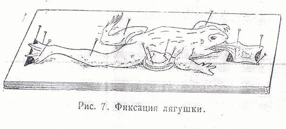
| The task № 3. | Study arterial hyperemia in frog’s tongue. |
Technique. Make a preparation of frog’s tongue as it is shown in the picture № 1. The received preparation of tongue examine under a microscope at small increase. Study an initial condition of a blood-flow in micro vessels. Then, lift an objective of a microscope and not changing position of a frog; slightly paint the tongue with cotton ball, moistened with solution of sodium chloride.
Lower a tube of a microscope on a former place, study changes of a blood-flow in micro vessels of frog’s tongue. Mark microscopic symptoms of arterial hyperemia.

|
| The task № 4. | Study venous hyperemia in frog’s tongue. |
Technique. Use the same preparation of frog’s tongue, as in the task № 4. Reproduce venous hyperemia by bandaging the basic vessels at a root of tongue. Separate the vein from an artery and a nerve, bring a tourniquet under the vein. The same manipulation make on the other side of a root of frog’s tongue. Under a microscope at small increase investigate blood circulation after bandaging of one vein then another. Draw the microscopic picture.
| The task № 5. | Receive fatty embolism and study blood circulation at embolisms of frog’s mesentery vessels. |
Technique: Open a thorax of unmoved frogs, release heart from a pericardium. Inject 0.2-0.3ml of warm petroleum jelly oil into heart ventricle with a thin needle. Under a microscope at small increase investigate blood circulation in mesentery vessels of the frog, fatty embolus movement and disorders of blood-flow caused by embolism.
Glossary
Arterial hyperemia is a local increased volume of arterial blood in tissue, resulting from augmented tissue inflow due to arteriolar dilation.
Артериальная гиперемия - увеличение кровенаполнения органа или участка ткани вследствие увеличения притока крови
Артериялық гиперемия – органға немесе тіннің бөлігіне қан ағып келуі көбеюінен қан толуының ұлғаю
Venous hyperemia (passive hyperemia, venous congestion) is a local increased volume of venous blood in a tissue resulting from impaired outflow from a tissue
Венозная гиперемия - увеличение кровенаполнения органа или участка ткани вследствие затруднения оттока крови
Веналық гиперемия – органнан немесе тіннің бөлігінен қан ағып кетуі қиындауынан оған қан толуының ұлғаюы
Ischemia is deficient blood supply to a part of tissue
Ишемия - уменьшение кровенаполнения органа или участка ткани вследствие уменьшения притока крови
Ишемия – органға немесе тіннің бөлігіне қан ағып келуі азаюынан оған қан толуының азаюы
Postischemic reperfusion is restoration of blood flow after ischemia
Постишемическая реперфузия-восстановление кровотока после ишемии
Ишемиядан кейінгі реперфузия – ишемиядан кейін қан ағысының қалпына келуі
Stasis is cessation of blood flow in capillaries
<емиядан кейін қан ағысының қалпына келуіStasis is cessation of blood flow in capillaries
Стаз - остановка кровотока в сосудах микроциркуляторного русла
Стаз – микроқанайналым арнасы қан тамырларында қан ағысының тоқтауы
Thrombosis is the lifetime clotting of blood in the lumen of the vessels or the cavities of the heart.
Тромбоз – прижизненное свертывание крови в просвете сосудов или полостях сердца.
Тромбоз – тірі адамның қан тамырында немесе жүрегінің қуысында қан қюы
Embolism is the circulation of particles, which normally do not occur in blood and obstruction of the vascular lumen
Эмболия – циркуляция в крови частиц, которые в норме не встречаются и закупорка ими просвета сосудов
Эмболия – қалыпты жағдайда кедеспейтін бөлшекткрдің қанменбірге айналымда болуы және олармен қан тамырларының бітелуі