The structures that support the teeth
Healthy teeth are, of course, embedded in bone. The bone is covered with gums, and the gums attach not only to the bone, but also to the tooth itself.
The gingiva is that portion of the gums that surrounds the teeth and lies above the level of the bone. The part of the gingiva below the crest but above the attachment is called the free gingival margin. The potential space between the free gingival margin and the tooth is called the gingival sulcus.
The periodontal Ligament (PDL)
The periodontal ligament is the soft tissue that lies between the tooth and its bony socket. It is a continuation of the connective tissue associated with the gingivo-dental fibers, and it continues around the entire tooth. In a healthy situation, there is never a direct attachment between the bone and the tooth itself. The PDL is composed of fibrous connective tissue. The bone that supports the teeth is called alveolar bone. It's only purpose in life is to support the teeth. The part of the alveolar bone that lines the socket is a thin layer of dense cortical bone called the lamina dura. The bone that underlies the lamina dura is cancellous bone (sometimes called medullary bone). Cancellous bone looks spongy and contains blood producing "organ" called bone marrow.
Notes:
| embedded [Im'bedId] вмурований, вставлений crest [krest] гребінь, виступ margin ['mQ:dZIn] край sulcus ['sulkəs] борозна ligament ['lIgqmqnt] зв’язка alveolar ['xlvIqlq, "xlvI'qVlq] альвеолярний | cortical ['kO:tIkl] кортикальний, корковий fiber['faIbq] волокно cancellous ['kxns(q)lqs] губчастий medulary [mq'dAlFrI] мозковий spongy ['spAndZI] губчастий bone marrow ['mxrou] кістковий мозок |
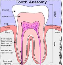
Ex.15. Have a look at the picture and speak about tooth anatomy: